Skin Laxity

Multiple causes contribute to increased skin laxity. These include: natural aging, genetics, diet, stress, lifestyle, sun exposure, weight fluctuations, exposure to environmental toxins, and smoking.

At Reszko Dermatology we use only the latest skin tightening technologies.  Our approach is a personalized combination of treatments to tighten, lift and often also contour the desired areas.

Our frequently used modalities include:

DERMAL FILLERS/COLLAGEN BIOSTIMULATION

Juvederm, Voluma, Volbella, Vollure, Restylane, Lyft, Silk, Belotero, Radiesse, Sculptra

  • Re-contours and lifts to restore lost volume and balance facial and body feature

FIBROBLAST THERAPY with PLASMA PEN

  • Plasma pen is the most advanced, non-invasive, skin lifting, skin tightening and rejuvenation device used to treat wrinkles, and sagging skin by stimulating production of collagen and elastin in the skin

MESOPUNCTURE

  • Bio-stimulatory peptides stimulate collagen to create volume in the skin and antioxidants and amino acids create an anti-inflammatory effect that encourages healing and optimal rejuvenation

MICRONEEDLING RADIOFREQUENCY (RF) VIVACE, GENIUS

  • Minimally invasive procedure that uses radiofrequency energy to precisely deliver heat into the deeper layers of the skin for new collagen stimulation. Ideal for fine wrinkling of the skin, lifting.

NEW SILHOUETTE THREAD LIFT

  • FDA-approved, minimally invasive, non-surgical equivalent of face lift
  • Ideal for the treatment of sagging jowls and nasolabial (face parentheses) and melolabial (marionette lines) folds

PRP (PLATELET RICH PLASMA)

  • Cutting-edge therapy that harnesses the body’s own growth factor-rich platelets, injected or micro-needled into the skin for optimum repair

ULTHERAPY

  • Non-invasive, no “down-time” ultrasound “face-lift” that stimulates new collagen and elastin synthesis deep within the skin’s structural support
  • The only device approved to lift sagging skin

Sun Protection

Sun exposure is the most preventable risk factor for all skin cancers, including melanoma as well as skin aging.

The following are Skin Cancer Foundation sun safety guidelines:

  • Seek the shade, especially between 10 AM and 4 PM
  • Do not burn
  • Avoid tanning and UV tanning booths
  • Cover up with clothing, including a broad-brimmed hat and UV-blocking sunglasses
  • Use a broad-spectrum sunscreen with an SPF of 30 or higher every day
  • Apply 1 ounce (2 tablespoons) of sunscreen to your entire body 30 minutes before going outside. Reapply every two hours or after swimming or excessive sweating.
  • Keep newborns out of the sun. Sunscreens should be used on babies over the age of six months.
  • Wear sun-protective clothing
  • Use extra caution near water, snow and sand as they reflect damaging rays
  • Get vitamin D safely through a healthy diet, including vitamin supplements
  • Examine your skin head to toe every month
  • See your doctor every year for a professional skin exam

For Every Day Sun Protection

Sunscreen should be worn every day, regardless of the weather or season. Sun exposure occurs all day long, through clouds, through glass windows of our homes and cars.

Limiting sun exposure, by wearing hats/gloves/sun protective clothing and avoiding exposure between 10am-3pm, is essential. You should wear sunscreen every day on exposed areas (i.e., face, neck, V-neck, chest, hands/forearms, and legs) for both skin cancer protection and defense against photo-aging.

SPF (Sun Protection Factor) is defined as a ratio of time in the sun required to sunburn with sunscreen on to the length of time in the sun required to burn without sunscreen.

  • SPF30: blocks 96.7% of ultraviolet radiation
  • SPF50: blocks 98% of ultraviolet radiation

Choose a daily sunscreen that is at least SPF30 (if you have dark skin) or SPF 40-50 (if you have light skin) and has both UVA and UVB (i.e. broad spectrum) coverage. If you have a personal or family history of skin cancer, or if you are often outdoors, consider using a sunscreen with higher than SPF 50.

There are two basic types of sunscreen:

  • Mineral sunscreen (physical blocker): physically blocks ultraviolet radiation. Active ingredients include zinc oxide or titanium dioxide.
  • Chemical sunscreens: contain special chemicals that absorb ultraviolet radiation such as oxybenzone, avobenzone, ecamsule, padimate O,PABA, octyl methoxycinnamate, octyl salicylate, phenylbenzimidazole sulfonic acid, homosalate, octisalate, octocrylene, octinoxate.

For face: UVA/UVB (broad spectrum) coverage higher than SPF30 is recommended

  • There are now many facial moisturizers that contain sunscreen, and this is adequate for regular daily use if the majority of your day is spent indoors and/or out of direct sunlight. Re-apply the sunscreen every few hours throughout the day.
  • Choose a non-comedogenic (non-pore clogging) sunscreen with zinc oxide and/or titanium dioxide if you have acne-prone skin or if you experience skin irritation with other chemical-containing sunscreens. Consult your doctor if sunscreens cause persistent significant skin irritation, or if you believe that you have a sunscreen allergy.
  • There are several powder-based products containing mineral sunscreens that can be applied over moisturizer or make-up.
    For lips: lip-balm containing SPF30 is recommended
    For body: UVA/UVB (broad spectrum) coverage greater than SPF50 is recommended
  • Lotion, cream, gel is recommended over spray.
  • Consider wearing SPF30-50 UV-blocking articles of clothing as an alternative to sunscreen.

What about sun protection for sports and outdoor activities?

For water sports, a wetsuit or UV-proof (SPF50) water jersey is recommended. Other sun-exposed areas require special sunscreens that will stay on in the water or after sweating. They need to be reapplied often, especially after sweating, toweling off or after extended periods of time when skin is immersed in water.

  • Very water resistant sunscreens: SPF is maintained after 80 minutes water immersion
  • Water resistant sunscreens: SPF is maintained after 40 minutes water immersion

Scars

Scar (also knows as cicatrix) is an area of fibrous tissue that replaces normal skin after injury.  Scar tissue is not identical to the tissue that it replaces and is usually of inferior functional quality. For example, scars in the skin are less resistant to ultraviolet (UV) radiation, and are devoid of sweat glands and hair follicles.

Scars, whether following skin surgery or trauma, are unpredictable. Many variables affect the final appearance of the scar.  These include the size and depth of the wound, the blood supply to the area, the thickness and color of your skin, the direction and the tension of the scar.

While no scar can be removed completely, the appearance of a scar can be improved and less noticeable through techniques described below.


SCAR & INJURY TREATMENT OPTIONS

    • Dermabrasion involves mechanical removal of the superficial layer of the skin (epidermis) and is most useful for the treatment of raised scars.   Dermabrasion is performed under local anesthesia.
    • Filler (hyaluronic acid, calcium hydroxyappetite) injections can be used to raise depressed scars to the level of surrounding skin. The effects are however temporary lasting on average 6-18 months.
    • Laser surgery and resurfacing – The use of lasers and light sources is the newest method of scar treatment. Over the past several years cosmetic resurfacing lasers have gained FDA approval for the treatment of scars. By removing superficial layers of skin resurfacing lasers may also help flatten scars.
    • Other cosmetic lasers including vascular lasers (pulsed dye laser, PDL) have been proven to greatly reduce the redness of most scars 6–10 weeks after the initial treatment.
    • Surgical scar revision – While no scar can be removed completely, the scar revision surgery may improve appearance of a scar. If you’re considering scar revision, the information below will give you a basic understanding of the most common types of scars and the procedures used to treat them.

Making the Decision

Many scars that appear large and unsightly at first may become less noticeable with time.  Scars mature and remodel becoming more conspicuous over the period of a year. For this reason, we tend to recommend waiting as long as a year or more after an injury or surgery before you decide to have scar revision.

Facial Scars

Because of its location and high visibility, a facial scar is frequently considered a cosmetic problem.  There are several ways to make a facial scar less noticeable. Often it is simply cut out and closed with small plastic surgery stitches, leaving a thinner, less noticeable scar.

If the scar lies across the natural skin creases, we may be able to reposition it to run parallel to these lines, where it will be less conspicuous.

Z-Plasty

Z-plasty is a surgical technique used to reposition a scar so that it more closely conforms to the natural lines and creases of the skin, where it will be less noticeable. It can also relieve the tension caused by scar contracture.

In this procedure, the old scar is removed and new incisions are made on each side, creating small triangular flaps of skin. These flaps are then rearranged to cover the wound at a different angle, giving the scar a “Z”pattern. The wound is closed with fine stitches, which are removed a few days later. Z-plasty is usually performed as an outpatient procedure under local anesthesia.

Contracted Scars

Burns or other injuries resulting in the loss of a large area of skin may form a scar that pulls the edges of the skin together, a process called contraction. The resulting contracture may affect the adjacent muscles and tendons, restricting normal movement.

Correcting a contracture usually involves cutting out the scar and replacing it with a skin graft or a flap. In some cases Z-plasty may be used.  In larger and complex cases newer techniques, such as tissue expansion, are playing an increasingly important role. If the contracture has existed for some time, physical therapy after surgery may be necessary to restore full function.

Skin Grafting and Flap Surgery

Skin grafts and flaps are more involved than other forms of scar surgery. Grafting involves the transfer of skin from a healthy part of the body to cover the injured area. The graft is said to “take “when new blood vessels and scar tissue form in the injured area.

After Scar Revision

With any kind or scar revision, it’s very important to follow your post-op instructions to ensure that the wound heals properly. As you heal, keep in mind that no scar can be removed completely; the degree of improvement depends on the size and direction of your scar, the nature and quality of your skin, and how well you care for the wound after the operation.

Radiotherapy

Radiotherapy is a reserved for the treatment of severe keloids and hypertrophic scars.  Low-dose, superficial radiation therapy, is used to prevent re-occurrence of severe keloids and hypertrophic scarring. It is usually effective, but only used in extreme cases due to the risk of long-term side effects.

Vitamins

Vitamin C normalizes collagen production and encourages the production of an organized, healthy collagen framework which improves scar appearance of the scar.  Vitamin C and some of its esters also fade the dark pigment associated with some scars.

Research shows the use of vitamin E and onion extract as a treatment for scars is ineffective. Vitamin E causes contact dermatitis in up to 33% of users and in some cases it may worsen scar appearance.


Abnormal Scars

Two types of scars are the result of the body overproducing collagen, which causes the scar to be raised above the surrounding skin.

A HYPERTROPHIC SCAR is an overgrown scar that remains within the confines of the initial injury or cut.  Hypertrophic scars often improve in appearance after a few years.  Hypertrophic scars are usually present without symptoms, although they might be itchy, red and relatively firm.They often improve on their own-though it may take a year or more-or with the help of steroid applications or injections.

If a conservative approach doesn’t appear to be effective, hypertrophic scars can often be improved surgically. Excess scar tissue is then removed and incision lines are repositioned in a less visible pattern. This surgery may be done under local or general anesthesia, depending on the scar’s location. You may receive steroid injections during surgery and at intervals for up to two years afterward to prevent the thick scar from reforming.


Keloid

A Keloid is a large, irregular scar that outgrows the site of initial skin injury. The tendency toward the development of hypertrophic scars may be inherited. Keloid scars can occur on anyone, but they are most common in dark-skinned people. Keloids can appear anywhere on the body, but they’re most common over the breastbone, on the earlobes, and on the shoulders. The tendency to develop keloids lessens with age. Keloids are often treated by injecting a steroid medication directly into the scar tissue to reduce redness, itching, and burning. In some cases, this will also shrink the scar.

If steroid treatment is inadequate, the scar tissue can be cut out and the wound closed.   This is generally an outpatient procedure, performed under local anesthesia. A skin graft is occasionally used, although the site from which the graft was taken may then develop a keloid.

No matter what approach is taken, keloids have a tendency to recur, sometimes even larger than before. To discourage this, we may combine the scar removal with steroid injections, direct application of steroids during surgery, or radiation therapy. Or you may be asked to wear a pressure garment over the area for as long as a year. Even so, the keloid may return, requiring repeated procedures every few years.

Surgical excision of hypertrophic or keloid scars is often associated to other methods such as presso-therapy or silicone gel sheeting. Excision of keloid scars without any adjuvant therapy shows a high recurrence rate close to 45%.  


Clinical Features of Hypertrophic Scars and Keloids

Hypertrophic Scars                                                                        Keloids

Develop soon after skin trauma/surgery                                   May develop months after the trauma

Usually improve with time                                                           Rarely improve with time

Remain within the confines of the wound                                Spread outside the wound

May improve with appropriate surgery                                     May worsen by surgery

Have no association with skin color                                           Are associated with darker skin color

Rosacea

Rosacea is a common, chronic skin disorder that frequently affects light-skinned Caucasian population.  Overall prevalence of rosacea ranges from less than 1% to 10%. The nose, cheeks, chin, forehead, and glabella are the most frequently affected sites. The disease has a variety of clinical manifestations ranging from flushing, persistent erythema, telangiectasias, papules, pustules and sebaceous gland hyperplasia.

Lasers and light sources revolutionized the treatment of rosacea.

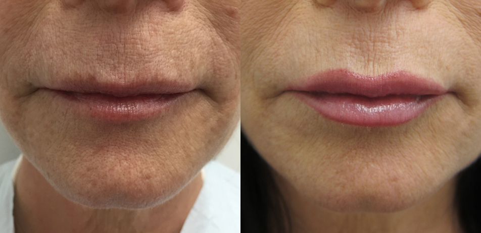
GLOBAL SKIN REJUVENATION with Aerolase

  • Concern: uneven skin tone, redness, skin inflammation, papules, pustules
  • Solution: Aerolase® (1064 Nd:Yag 650 microsecond) targets redness and melanin within the skin. In addition to improvement of rosacea redness and skin sensitivity, patients you can see an overall improvement in skin tone, texture, and elasticity.
  • Expectations: Patients can expect mild warming sensation with little to no post-procedural downtime (~few hours-days of redness). 3-4 treatments are recommended for optimal results

IPL (INTENSE PULSED LIGHT) 

  • Concern: irregular or unwanted rosacea redness
  • Solution: IPL is light-based therapy targets redness, broken capillaries for a clear, radiant, even-toned complexion.
  • Expectations: Patients should expect a mild warming sensation during treatment with minimal to no downtime. Around 6 treatment sessions are recommended for rosacea redness.

MICRONEEDLING RADIOFREQUENCY (RF) VIVACE, GENIUS 

  • Concern: surface irregularities, redness, skin sensitivity
  • Solution: Vivace and Genius combine radiofrequency and gold-plated microneedling technology, delivering heat into the deeper layers of the skin promote new collagen rich tissue, which helps to even skin tone, while also helping to lift and tighten the skin. Vivace and Genius can help globally enhance the appearance of skin and reduce surface irregularities present in rosacea.
  • Expectations: Following local anesthesia in the treatment areas, most find the treatment very tolerable with little to no downtime. 2-4 treatments are recommended for optimal collagen induction.

PERFECTA V-BEAM VASCULAR LASER

  • Concern: dilated or broken capillaries, overall redness
  • Solution: This laser produces a burst of light to specifically target vascular lesions, unwanted blood vessels, and redness that are common in rosacea.
  • Expectations: Patients should expect a mild warming sensation with no local anesthesia required. 3-4 treatments are best for optimal reduction of rosacea redness.

Currently, only the following medications are FDA-approved for use in rosacea:

  1. Topical medications: metronidazole (0.75 and 1%) (gel, cream, lotion), azelaic acid (15 and 20%) cream, sulfacetamide 10%-sulfur 5% (gel, cream, topical suspension, wash)
  2. Systemic: Oracea 30 mg immediate release & 10 mg delayed release doxycycline

Other non-FDA approved treatments used off-label have also been beneficial and will be discussed below.

Extended information on rosacea

Rosacea is a common, chronic skin disorder that frequently affects light-skinned Caucasian population.  Overall prevalence of rosacea ranges from less than 1% to 10%.  The nose, cheeks, chin, forehead, and glabella are the most frequently affected sites.  The disease has a variety of clinical manifestations ranging from flushing, persistent erythema, telangiectasias, papules, pustules and sebaceous gland hyperplasia.  Significant psychological stress and diminished perceive quality of life often accompanies this condition.  The pathogenesis is likely multifactorial and includes genetic and vascular elements, climatic exposures, pilosebaceous unit abnormalities, and possibly microbial organisms.

Diagnosis of rosacea is based primarily on clinically recognizable morphologic characteristics. An expert committee assembled by the National Rosacea Society on the Classification and Staging of Rosacea defined and classified rosacea in April 2002 into 4 clinical subtypes based primarily on morphologic characteristics.  The subtypes include erythemato-telangiectatic rosacea (ETR), papulo-pustular rosacea (PR), phymatous rosacea, and ocular rosacea.

In order to design appropriate treatment regimen it is essential to correctly identify the subtype of rosacea.   In addition, rosacea has to be distinguished from its clinical mimickers.  ETR must be differentiated from chronic sun damage and photodermatitis.PR must be distinguished from acne vulgaris, seborrheic dermatitis, lupus miliaris disseminates faciei, collagen vascular diseases, perioral dermatitis, and Demodex folliculitis.  Differential diagnosis of ocular rosacea includes allergic conjunctivitis and blepharitis.

Therapy of rosacea is centered on symptomatic improvement i.e. reduction of facial erythema and number of inflammatory lesions, decrease in the number, duration and intensity of flares, and reduction of concomitant symptoms of itching, burning, and facial tenderness.

In addition to medical therapy sun protection and appropriate skin care regiment need to chosen as rosacea patients often exhibit marked skin sensitivity and suffer from intolerance to skin products and cosmetics. Triggers for cutaneous flushing should be identified and eliminated.   Commonly identifiable triggers include topical cosmetics and corticosteroids, alcohol, tobacco, exercise, hormonal changes, sun exposure, hot weather, exercise, ingestion of hot or spicy foods/ drink, caffeine, emotional stress and medications such as topical and systemic steroids, niacin and nitroglycerin. A flare of rosacea may be anticipated when steroids are discontinued and may be dramatic requiring therapy with systemic antibiotics and topical tacrolimus.

Currently, only the following medications are FDA-approved for use in rosacea:

  • Topical medications: metronidazole (0.75 and 1%) (gel, cream, lotion), azelaic acid (15 and 20%) cream, sulfacetamide 10%-sulfur 5% (gel, cream, topical suspension, wash)
  • Systemic: Oracea 30 mg immediate release & 10 mg delayed release doxycycline

Other non-FDA approved treatments used off-label have also been beneficial and will be discussed below.

Erythemato-telangiectatic rosacea (ETR)

ETR subtype is clinically characterized by diffuse erythema and telangiectasias on the cheeks, forehead, dorsal nose, or sometimes entire face.  Patients frequently complain of intolerance or sensitivity to topical products and cosmetics.

This type of rosacea is best treated with avoidance of triggers leading to flushing, strict photoprotection, electrosurgery and phototherapy.  In recent years phototherapy and laser therapies are gaining popularity as treatments of rosacea associated erythema and telangiectasias.  Laser therapies include pulse-dye laser (PDL), potassium-titanyl-phosphate (KTP) laser, Nd-YAG laser, or intense-pulsed light (IPL).  In a study of 34 patients with ETR, IPL treatment resulted in overall patient and physician reported over 50% improvement in 73% and 83% of patients, respectively. The results were sustained at 6 months.  In a different study 83% of patients had less facial redness, 75% noted less flushing and better skin texture, and 64% noted fewer acneiform breakouts.

Topical selective alpha-agonist, oxymetazoline, represents a novel approach to management of ETR associated erythema and flushing.   A small study showed significant benefit with the use of the alpha-agonist oxymetazoline in reducing the flares and the inflammatory symptoms in patients with traditional medication resistant ETR.

Facial edema may be a prominent clinical feature of rosacea.  Recurrent vasodilation results in a feeling of fullness of the cheeks and visible subtle induration of the cheeks, so called solid facial edema.   Solid facial edema of ETR responds to isotretinoin.

Papulo-pustular rosacea (PR)

PR subtype is characterized by papules and pustules often on erythematous base primarily affecting the nose, cheeks, and forehead. Predilection of the lesions on the central, convex aspect of the face, sometimes with corresponding central facial edema is frequently noted.

Topical Therapy

The major topical antibiotics used to treat PR are metronidazole, clindamycin and erythromycin. Other topical therapies include azelaic acid, pimecrolimus, antiparasitics, alpha-adrenergic agonists and topical sulfacetamide 10% with sulfur 5%.

Metronidazole

Clinical efficacy of the topical metronidazole in the treatment of rosacea has shown in a number of clinical trials.  The mechanism of action of metronidazole is not yet well established, but appears to be anti-inflammatory.  Topical metronidazole is commercially available in a 0.75% gel, lotion and cream for twice daily use, and a 1% cream or gel for once daily use.   Twice-daily 0.75% metronidazole was shown to be well-tolerated and effective in the treatment of 582 patients with mild to moderate severity PR.  Mean erythema severity score was reduced by 50% by week 12 of treatment. In a 12-week, randomized control study, Jorizzo et al. showed that once daily dosing of 1% metronidazole cream is as effective as twice daily dosing.  Metronidazole is generally well tolerated and has a low incidence of adverse side effects.  Most commonly reported include mild pruritus, skin irritation and dryness.

In a recent Cochrane review, topical metronidazole was shown to be significantly more effective than a vehicle in 174 patients with marked reduction of number of inflammatory lesions and erythema scores with an odds ratio of 5.96.  Metronidazole plays a role in maintenance therapy, either with or without prior concomitant systemic antibiotic therapy. Effect of metronidazole on rosacea associated telangiectasias is generally minimal.

Sodium sulphacetamide with sulphur

Despite the lack of randomized controlled studies, sodium sulphacetamide 10% with sulfur 5% has been used as a safe, well-tolerated and effective treatment option for rosacea for over 50 years.  Eight week therapy with sodium sulphacetamide 10% with sulfur 5% resulted in a significant reduction in inflammatory lesions and facial erythema (78% vs. 36% and 83% vs. 31%, respectively) compared to a vehicle.  Adverse effects of topical application were generally mild and included pruritus, contact dermatitis, irritation, erythema, scale and xerosis.

Newer, emollient foam formulation of 10% sodium sulphacetamide and 5% sulfur offer similar benefits with less lingering odor and reduced irritation potential. Preliminary anecdotal evidence suggests synergistic benefit of concurrent sodium sulphacetamide10% with sulfur 5% and metronidazole application.

Azelaic acid

Azelaic acid, commercially available as 15% gel or 20% cream, is a naturally occurring, saturated 9-carbon dicarboxylic acid derived from Pityrosporum ovale.  Azelaic acid is FDA-approved for the treatment of mild to moderate rosacea.  Its biologic effects encompass anti-inflammatory, anti-keratinizing and anti-bacterial properties.  Recently, PPAR gamma activation was shown as a main modulator of the inflammatory response modulated by azelaic acid in normal human keratinocytes.

Clinical safety and efficacy of azelaic acid 15% gel applied twice daily for 12 weeks has been demonstrates in two phase III, vehicle-controlled, randomized trials of 664 patients with PR. On average improvement of erythema ranged from 44-46% in patients treated with azelaic acid, compared with 28-29% in the vehicle group.   A mean reduction in inflammatory papules and pustules ranged from 51-58% in the azelaic acid group, compared with 39-40% in the vehicle group.

Data from three clinical trials analyzed by Cochrane review showed rates of improvement in azelaic acid group of 70-80% compared with 50-55% in the placebo group.  In a randomized trial comparing topical metronidazole with azelaic acid the efficacy of 20% azelaic acid was greater than metronidazole in improving erythema and inflammation in physician rating of global improvement.  Application of azelaic acid was associated with a greater irritation.  Wolf et al. found the efficacy of once-daily application of metronidazole 1% gel and twice daily azelaic acid 15% gel to be similar.  Both medications were well tolerated, with the number of adverse effects slightly higher in azelaic acid group.  All reported adverse effects were mild to moderate.

Anecdotal evidence suggests beneficial effects of topical clindamycin, erythromycin, and combination benzoyl peroxide-clindamycin in the treatment of rosacea, but efficacy of these modalities have not yet been evaluated in well-designed clinical trials.  In a double-blind, randomized controlled trial of once daily clindamycin 1% benzoyl peroxide 5% gel a significant reduction in inflammatory lesion count and erythema was noted. The treatment was generally well tolerated.

Tacrolimus and pimecrolimus

Topical tacrolimus (0.03% or 0.1%ointment) and pimecrolimus (1% cream) are macrolide non-steroidal immunomodulators.  Their mechanism of action is by inhibition of T-cell activation and inhibition of subsequent cytokine release.  In a pilot study, topical pimecrolimus showed clinical benefit with decreased erythema and number of inflammatory lesions in after 12-18 weeks of therapy.  Another randomized trial, nonetheless, demonstrated that pimecrolimus was no more effective than the vehicle when used for 4 to 6 weeks.

A recent single-centre, randomized, open-label study of 48 patients comparing pimecrolimus with metrondazole both treatments were very effective in the treatment of PR. There were no significant differences between the treatments in inflammatory lesion counts, overall erythema severity scores and physician global assessment scores evaluated from baseline to week 12.  Neither treatment produced any clinically relevant improvement in telangiectasia.

Rarely pimecrolimus may produce a syndrome similar to steroid-induced acne.

Anecdotal evidence suggests that topical tacrolimus may be an effective treatment of steroid induced rosacea especially when combined with systemic minocycline.

Retinoids

The role of topical retinoids in the management of rosacea remains controversial.  The mechanism of action of the retinoids is by regulation of the retinoic acid receptor, an important regulator of keratinocyte proliferation, differentiation, and cutaneous inflammation.  Use of retinoids carries a theoretical benefit of decreased angiogenesis and decrease in cutaneous vascularity and possible prevention of new telangiectasias.  In a few small series, topical retinoid, tretinoin, have demonstrates benefit for rosacea with a lessening degree of erythema and a partial to complete disappearance of telangiectasias as well as decreased in the number of inflammatory lesions although the clinical response was delayed and not evident until 2 or more months of therapy.

A small study demonstrated the efficacy of topical free radical scavenger vitamin C for rosacea suggesting the possibility that free radical production may play a significant role in the pathological inflammatory response seen in rosacea.

Some antiparasitic drugs have been observed to be helpful as both oral and topical agents in the treatment of the inflammatory rosacea. Oral ivermectin and lindane have shown some efficacy in the eradication of Demodex folliculorum, one possible factor that may lead to the inflammation underlying rosacea.  Permethrin and crotamiton topically have not shown great usefulness in eradicating Demodex folliculorum, but may still lead to the resolution of some of the underlying inflammation. In a small randomized double-blind placebo-controlled study permethrin 5% cream was found superior to the vehicle and similar in efficacy to 0.75% metronidazole gel.


Oral Therapy

Antibiotics

Tetracyclines are broad-spectrum antibiotics that remain the mainstay of oral pharmacotherapy for rosacea.  Tetracycline (250 to 500 mg twice daily) and more recently second generation tetracyclines, doxycycline (100 to 200 mg per day and extended release 20-40 mg per day) and minocycline (100-200 mg per day),are commonly used oral therapies.  Second generation tetracyclines have an improved bioavailability, longer elimination half-life, once daily dosing and can be taken with food which minimizes gastrointestinal side effects.  Azithromycin has also been shown to be beneficial for treating rosacea, is better tolerated than first generation tetracyclines and can be dosed 3 times a week.

The precise mechanism of action of tetracyclines remains unknown.  Most likely anti-inflammatory and not anti-microbial actions of tetracyclines such as their actions on inhibition of angiogenesis, neutrophil chemotaxis, release of pro-inflammatory cytokines, cellular apoptosis, cell proliferation and inhibition of matrix metalloproteinases contribute to clinical benefit in management of rosacea.   A significant benefit of second generation tetracyclines is their clinical effectiveness at sub anti-microbial, anti-inflammatory doses limiting undesired side effects (candidal vulvovaginitis, gastrointestinal distress) and possibly bacterial resistance. Systemic tetracycline antibiotics should not be used in children under the age of 8 because of potential permanent discoloration of teeth.

Anti-inflammatory dose of doxycycline, a 40 mg doxycycline monohydrate containing 30 mg immediate-release and 10 mg delayed release doxycycline, commercially available as Oracea, is the only tetracycline approved in the US for the long-term (12 months) use.  Oracea has been shown effective in treatment of PR with favorable risk/benefit ratio.  Used at sub anti-microbial doses, long-term use of anti-inflammatory doxycycline might not exert selective pressure on bacteria, and thus limit development of bacterial resistance.

Two phase III, controlled studies have demonstrated the safety and efficacy of a 16-week treatment with anti-inflammatory 40 mg doxycycline administered daily in the management of PR. Both studies included patients with inflammatory PR with 10-40 papules, moderate to severe erythema and telangiectasias.  At the end of 16 weeks, the mean change in lesion count in doxycycline groups was -11.8 and -9.5 in compared with -5.9 and -4.3 in the placebo group?  Doxycycline was well tolerated with low incidence of side effects.  Most commonly reported side effects included nasopharyngitis, diarrhea and headaches.  Efficacy beyond 16 weeks and safety beyond 9 months has not yet been established in randomized controlled studies.

Current research on small number of patients suggests that combination therapy of oral anti-inflammatory dose of doxycycline and topical metronidazole leads to quicker and more effective alleviation of inflammatory lesions.

Oral erythromycin at 250-1000 mg per day is considered an effective drug for the treatment of PR.  The use of erythromycin is reserved for those patients that are intolerant, allergic or refractory to tetracyclines or in cases when tetracycline therapy is contraindicated (e.g. pregnancy).

Second generation macrolides, clarithromycin and azithromycin have both been found to be effective and tolerable drugs in short term therapy of rosacea.  At the end of a 12-weektreamtent with azithromycin, a 75% decrease in total scores and an 89% decrease in inflammatory lesion scored was noted compared with baseline values.

A significant amount of circumstantial evidence links the eradication of Helicobacter pylori with the “triple therapy,” consisting of omeprazole and 2 of the following antibiotics: clarithromycin, amoxicillin, or metronidazole and the successful treatment of rosacea.

Metronidazole

In a randomized study of oral metronidazole (200 mg twice daily) vs. oxytetracycline (250 mg twice daily) both drugs produced an improvement after 6 and 12 weeks of therapy with no significant differences between the two.   In rare instances, treatment with oral metronidazole might be associated with epileptiform seizures, encephalopathy, and sensory neuropathy.  Oral metronidazole also requires abstinence from alcohol.

Isotretinoin

In a small number of clinical trials, isotretinoin was found to be effective for both ETR and PR.  Isotretinoin, although not working directly as an antibiotic, has strong antibacterial effects.  Irvine et al. noted a reduced facial cutaneous blood flow in the isotretinoin group compared with oxytetracycline group.

A small study of 22 patients showed the effectiveness of a 4 month low-dose (10 mg) isotretinoin therapy in patients with therapy-resistant rosacea.  The treatment led to a reduction of number of inflammatory lesions, erythema and telangiectasia.   The overall effect of isotretinoin therapy was delayed in comparison to therapy with oral antibiotics.

Gollnick et al. compared efficacy of isotretinoin (0.1. mg/kg, 0.3 mg/kg or 0.5 mg/kg) with doxycycline (100 mg daily for 14 days, followed by 50 mg daily) in the treatment of grade II and III rosacea in a double-blinded randomized 12 week study.  Isotretinoin 0.3 mg/kg proved to be the most effective dose compared with placebo. Isotretinoin 0.3 mg/kg treatment resulted in 90 % reduction of number of lesions compared with 83 % reduction with doxycycline.  Isotretinoin 0.3 mg/kg was generally well-tolerated.

Other oral therapies

Other oral therapies that have shown benefit are medications that reduce flushing beta-blockers, clonidine, naloxone, ondansetron, and selective serotonin reuptake inhibitors.

Oral contraceptive therapy has been helpful in patients who provide historical information of worsening rosacea with their hormonal cycle.

Dapsone has been used in severe, refractory rosacea, and dapsone has been particularly beneficial for patients who cannot take isotretinoin.


Phymatous rosacea

Phymatous rosacea is characterized by marked by thickening of the skin, irregular skin texture, edema, hypertrophy and hyperplasia of sebaceous glands, connective tissue, and vascular bed.  Phymatous rosacea commonly involves the nose (rhinophyma) but changes can also be seen on the chin (gnathophyma), ears (otophyma), forehead (metophyma), and eyelids (blepharophyma).  This severe form of rosacea requires aggressive medical therapy with systemic antibiotics, and sometimes warrants use of isotretinoin. Once the medical regimen has been maximized, the patient is a candidate for surgical/ laser resurfacing of the affected area.  Electrosurgical sculpturing of the phymatous skin gives excellent results, is fast, and is almost bloodless.   A CO2 laser or erbium-YAG laser may also be used for the treatment of rhinophyma, although it is more costly and time-consuming than electrosurgical treatment. Electrosurgical treatment is more likely to scar compared to laser therapy.

Cold-steel surgery and dermabrasion are also effective, but they are more difficult owing to the vascularity of the nose.


Ocular rosacea

Ocular rosacea can be seen in the presence or absence of skin manifestations of rosacea. Clinically, ocular rosacea is characterized by conjunctival erythema and injection, sometimes accompanied by eyelid edema), foreign body sensation, inflammation of the lids , blepharitis and meibomian glands, interpalpebral conjunctival hyperemia, conjunctival telangiectasias and/or glandular inflammation (chalazion) along the eyelid margin.43  Patients report subjective symptoms: foreign body sensations, dry eyes, stinging, itching and burning, photosensitivity. The vision is rarely affected. Ocular rosacea requires systemic antibiotics, and oral tetracyclines including low dose doxycycline are an excellent first-line therapy for this form of rosacea.  Topical corticosteroid eyedrops may also be beneficial.


Other rosacea subtypes

Granulomatous rosacea is a variant of rosacea clinically presenting with erythemaotus to brownish papules with peripheral erythema on the malar and perioral region.  Treatment of granulomatous variant is difficult.  In a case report, granulomatous rosacea refractory to traditional treatments was treated with IPL with satisfactory improvement.44  Authors postulated that IPL system with non-fixed wavelengths, ranging from visible to infrared might provide added benefit of treating telangiectasias at different depths leading to subsequent improvement.

Rosacea fulminans (sudden onset of coalescent papules, pustules, nodules) may occur during pregnancy, thyroid diseases, depression, emotional stress, induced by medications.  This exacerbation may require use of systemic corticosteroids (prednisone 0.5 to 1 mg/kg/day) and/or isotretinoin.

Conclusion

The mainstay of rosacea therapy remains topical metronidazole or azelaic acid and oral tetracyclines. As this disease process causes increased skin sensitivity, many of the topical therapies may not be well tolerated, necessitating other therapies. It is therefore important to have as many treatment options as possible.

Pre-cancerous Skin lesions

Actinic keratoses (AKs) are common cutaneous lesions that occur on sun-exposed areas particularly in blond or red-haired, fair-skinned individuals with green or blue eyes. They of course can occur in patients that do not possess these phenotypic features and relate most directly to cumulative sun exposure.  AKs represent the initial intraepidermal manifestation of abnormal proliferation of keratinocytes with the possibility of progression to squamous cell carcinoma in situ (SCC in situ) and invasive squamous cell carcinoma (SCC).

Clinically, AKs have three possible behavioral patterns: spontaneous regression, persistence, or progression into invasive SCC.   The risk of progression of AK to SCC has been calculated to be between 0.025 and 16% per year and the calculated lifetime risk of malignant transformation for a patient with AKs followed for a period of 10 years ranges from 6.1 to 10.2%.  The long-term risk of development of invasive SCC in patients with multiple AKs has been estimated to be as high as 10%.  Approximately 60-65% of SCCs arise from prior AKs.  Spontaneous regression has been reported in as high as 25.9% of AKs over a 12-month period, although a 15% recurrence rate was noted at follow-up.

Pathogenesis

Factors linked to pathogenesis of AKs include

  • exposure to ultraviolet B (UVB) light; UVB causes mutations of the tumor suppressor gene (TSG) p53
  • genetic DNA instability (i.e. xeroderma pigmentosum) or melanin deficiency (albinism)
  • older age
  • male gender
  • anatomical location; over 80% of AKs are located on the head and neck, dorsal forearms and hands
  • history of immunosuppression; solid organ transplant patients are at significantly increased risk.

Clinical Features

AKs are red, pink, or brown papules with a scaly (hyperkeratotic) surface.  They occur on sun-exposed areas and are especially common on the balding scalp, forehead, face, dorsal forearms and hands.   Subclinical (non-visible) AKs are estimated to occur up to 10 times more often than clinically visible AKs, particularly on sun-exposed skin.

Treatment

Prevention of disease is always superior and preferable to the need for treatment. Effective preventative measures include avoidance of excessive sun exposure (use of broad-brimmed hats, sun-protective clothing and sunscreen), patient education, and regular self-examinations to detect the earliest signs of malignant transformation.

Due to their low but real potential to develop into invasive SCC, AK therapy is generally recommended.  The management options for AKs should be chosen depending on whether a lesion-directed or a field-directed therapy is preferred.  Lesion-directed therapy, including ablative and surgical procedures, is reserved for selected cases where only a few clinically visible AKs are present.  Field-directed therapy, including ablative, non-ablative and topical treatments, offers the advantage of treating both clinically evident and subclinical lesions that may progress to visible AKs.

Lesion-directed treatment

Cryotherapy is the most commonly used lesion-directed treatment modality.  Clearance rates range from 39 to 98.8%.  In a large, multi-center Australian study evaluating the efficacy of cryotherapy for the treatment of AKs on the face and scalp, of the 89 patients and 421 lesions in the intended-to-treat population, there was an average of 67.2% lesion response rate per patient.  Cryotherapy treatment was associated with “good” and “excellent” cosmetic outcomes in 94% of the lesions.

Other lesion-directed treatment options include electrodessication and curettage (ED&C) and surgical excision.  Treatment with ED&C may require two or three cycles.  High cure rates and good cosmetic outcome have been reported.  C&D should be generally avoided in recurrent lesions, punch-biopsied lesions and hair bearing sites.

Field-directed treatment

Field-directed treatment modalities include topical pharmacologic therapies including imiquimod, 5-FU and diclofenac, ablative and nonablative laser resurfacing, PDT, dermabrasion and deep and medium-depth chemical peels.

Retinoids

In a double-blind, randomized clinical trial 25 patients applied topical all-trans-retinoic acid (tretinoin) 0.05% cream twice daily for 16 weeks resulting in a 30.3% reduction in the number of AKs compared with baseline.

 Imiquimod (Aldara, Zyclara)

The manufacturer-recommended standard imiquimod treatment protocol for AKs is twice weekly for sixteen weeks.  Other treatment protocols (three times a week for four weeks followed by a 4 week rest and an additional 4 weeks of treatment if remaining lesions are still present; two to three times a week for 16 weeks) have been reported.  Clearance rates with imiquimod range from 45 to 85%.  Reported recurrence rates are 10 and 16% within 1 year and 18 months of treatment respectively.

Common reported side effects of imiquimod therapy included site reactions (edema, erythema, vesicles and erosions/ulcerations), fatigue, headache, diarrhea, nausea and leucopenia.  No serious adverse events were reported.

5-FU (Efudex and Carac)

Manufacturer-recommended 5-FU treatment protocol for AKs is twice daily for 2-4 weeks.  Other protocols proposed in the literature include 5% 5-FU (Efudex) once to twice daily for 2-7 weeks; 5% 5-FU once daily, one day per week for 12 weeks; and 0.5% 5-FU (Carac) once or twice daily for 1-4 weeks.  The literature reported rates of AK resolution with 5-FU that reach 89% with twice weekly application for 16 weeks, albeit recurrence rates of up to 55% have been reported.  In addition, low compliance, due to adverse effects of medication, is associated with significant treatment failure rates.

Diclofenac

Efficacy of topically applied 3% diclofenac gel in 2.5% hyaluronic acid vehicle gel vs. vehicle was addressed in a multicenter, randomized, double-blind, placebo-controlled study of 195 patients.  Treatment with 3% diclofenac gel twice daily for 30-90 days resulted in an improvement of 59% compared with 31% with placebo.

Photodynamic Therapy (PDT) (LINK)

PDT with ALA (dALA or δ-ALA or 5ala or 5-aminolevulinic acid) activated by either blue or red light showed reported overall clearance values for AKs of 80% and 60%, respectively.   Response rates vary with duration of ALA incubation and thickness of AK lesions.  In a study of 243 patients with total of 1403 AKs on scalp and face, were treated with ALA (incubation time 1-18 hrs) and exposure to blue light.  At 8 weeks, 30% of patients with partial response were retreated.  At 12 weeks, 91% lesion clearance rate was reported.  Discomfort was the most commonly reported adverse event. Satisfaction with the cosmetic outcomes for PDT was 96%.

Melasma

Melasma is a skin pigmentary disorder affecting millions of patients worldwide. Melasma is characterized by patterns of patchy hyperpigmentation, usually on the cheeks, under the eye, upper lip, and forehead. Skin discoloration associated with melasma is due to an over accumulation of skin pigment melanin, is produced by pigment cells called melanocytes. Although etiology of melasma is largely unknown, it is understood that genetics plays the largest role. Melasma can be further exacerbated by UV exposure, heat and visible light.  Occurrence of melasma is often linked to hormonal triggers like oral contraceptive pills, IUDs, hormone therapy and pregnancy.

Melasma can be categorized as “epidermal” or “dermal”, or mixed.

In epidermal melasma melanin is present in the more superficial epidermal layers

In dermal melasma melanin is in the deeper layers of the skin and is typically visualized best under Wood’s lamp examination. This distinction is crucial because epidermal melasma responds more quickly to treatment.  Dermal melasma is much more difficult to treat.

Mixed melasma is a combination of epidermal and dermal melasma.  Melasma is often associated with increased vascularization ie. increased number of blood vessels, vessel size, and vessel density.

When treating pigmentary disorders it is important to be evaluated by a board-certified dermatologist to first accurately diagnose melasma as an underlying cause of hyperpigmentation and second to determine melasma subtype prior to pursuing any treatments or products as some treatments might significantly worsen melasma.


Treatment for Melasma

The management of melasma is often challenging, with partial responses and frequent relapses. A combination of therapies targeting multiple pathogenic elements, such as photodamage, inflammation, aberrant vascularity, and abnormal pigmentation, generally provides the best clinical outcomes. The best therapeutic armamentarium includes a variety of agents that

  • inhibit the biosynthesis of melanin
  • increase skin epidermal turnover and therefore removal of excess of melasma pigmentation
  • chemical peels and lasers that directly remove of excess melanin

Sun Protection

Sunscreens are essential to prevent progression of melasma due to UV exposure, visible light, and blue light from electronic devices. The best sunscreen for melasma is physical sunblock with SPF 30 or higher that contains zinc oxide or titanium dioxide as the active ingredient.

Lasers

NeoClear by Aerolase

The NeoClear by Aerolase® has proven to be effective against both dermal and epidermal melasma. This laser targets pigment/melanin itself, without causing any additional tissue damage and without any risk of post-inflammatory hyperpigmentation or pigmentation worsening. This makes 1064 lasers an ideal choice in melasma treatment, as many other treatments (including both topicals and other lasers) result in tissue inflammation. Also unlike other lasers, due to its 650 – microsecond technology the laser can effectively penetrate to the deeper layers of the skin ie. dermal layers treating dermal melasma and melasma associated increased vascularity  without risk of tissue damage.

Clear + Brilliant, Permea

Clear + Brilliant is a non-invasive laser that creates thousands of “micro injuries” to the epidermis. These micro-injuries trigger the body’s healing process, replacing damaged cells with new, healthy tissue.  Clear and Brilliant resurfacing also allows for optimal penetration of products that help to reduce pigmentary change.  Clear + Brilliant is the most gentle fractional resurfacing laser (treating up to 20% of the skin), which is optimal for those who are looking to minimize downtime or those who have milder cases of melasma. Multiple sessions are required to achieve clinical reduction of melasma.

Fraxel DUAL 1550/1927 Fractional Resurfacing

Fraxel is a non-ablative fractional resurfacing laser that creates microthermal injury zones in the skin to stimulate the body’s own healing and rejuvenating process.  The procedure targets a portion of tissue at a time, leaving the surrounding tissue enact. As this laser does not cause complete tissue damage, the laser itself can remove tissue containing melanin pigment. Fraxel is from the same family as Clear + Brilliant, however Fraxel targets a larger percentage of skin tissue (treating up to 40% of the skin). This is an optimal treatment option for patients who are comfortable with having more downtime as well as patients with a most severe melasma.

Coolpeel CO2 Resurfacing, Lumenis UltraPulse CO2 Ablative Resurfacing

Considered one of the most intensive resurfacing lasers, the CO2 laser is an ablative fractional laser. By comparison to the other lasers in this category, the CO2 laser removes via vaporization the outermost (epidermal) layers of skin cells, relying on the dermal tissue beneath to initiate a healing response. This again creates new, healthier tissue that is free of pigmentary changes or evidence of UV damage. These lasers not only target the melanin pigment in the epidermal skin layers, but can also target the pigment producing melanocytes. This laser is extremely efficacious, but requires the most significant amount of downtime. This laser is ideal for patients who are comfortable with extended periods of downtime as well as patients with stubborn, resistant melasma that has not responded with more conventional laser therapies.

Oral medications

Tranexamic acid- which has been shown to be effective in the treatment of melasma. The largest study on the use of oral tranexamic acid for treatment of melasma was a retrospective review of 561 melasma patients treated with tranexamic acid.  More than 90% of patients received prior treatment for their melasma, including bleaching creams and laser treatments. Among patients who received oral tranexamic acid over a 4-month period, 90% of patients demonstrated improvement in their melasma severity. Tranexamic acid has been shown to be safe but is not recommended for a long term use. It is also contraindicated in patients with personal or family history of clotting disorders, personal history of deep vein thrombosis (DVT), or patients who are currently pregnant or are preparing to conceive.


CHEMICAL PEELS

Chemical peeling involves the topical application of a chemical agent, wherein the desired outcome is a controlled regeneration of the skin and removal of excess pigmentation.

Cosmelan depigmenting solution by mesoestetic offers a professional depigmenting protocol designed to eliminate or reduce dark spots while unifying the skin tone and enhancing overall skin luminosity.

Topical skin care

There are many active ingredients used to improve existing hyperpigmentation of melasma. The most common and effective ones are hydroquinone (HQ), kojic acid, azelaic acid, ascorbic acid/Vitamin C, glycolic acid (AHA or alpha hydroxy acids), tranexamic acid, licorice root extract, and Vitamin A derivatives (ie retinol, all-trans retinoic acid). Most of these ingredients work mostly by either inhibiting melanin synthesis or exfoliating excess of superficial pigmentation.

  • HQ- Of the components mentioned above HQ has shown to be one of the most effective topical treatments and is widely considered the gold standard in clinically reducing the appearance of melasma. However, due to its strong effects on skin cells, HQ should not be used for long periods of time, and the recommended duration should not exceed more than 3-6 months of continuous use.
  • Vitamin A (Retinoids, Retinols) – Acts by tyrosinase inhibition, preventing melanosome transfer, and increases epidermal turnover, by essentially “ungluing” the bonds that hold damaged, melanin-containing skin cells and effectively exfoliate these dead skin cells. Cellular turnover by using vitamin A is typically seen over a period of 28 days, however increased use of vitamin A has shown to increase the rate of cell turnover to 21 days or even 18 days. Vitamin A on its own and the combination of vitamin A + HQ can be very irritating and drying on the skin, so these medications are often compounded further with a corticosteroid (commonly called triple combination or TC therapy) to help reduce inflammation resulting from these active ingredients. They also make your skin increasingly photosensitive, which further stresses the importance of daily sunscreen with SPF of 30 or higher.
  • Vitamin B3 (Niacinamide) – Studies have shown that Vitamin B3 may decrease skin pigmentation by preventing melanosome transfer within skin cells. It also plays a role as both an antioxidant and an anti-inflammatory. Similarly to Vitamin C, Vitamin B3 neutralizes reactive oxygen species (ROS). As an anti-inflammatory, Vitamin B3 can effectively reduce the inflammatory response from intensive active ingredients such as HQ and Vitamin A.
  • Vitamin C (Ascorbic Acid) – Another tyrosinase inhibitor, widely used in skincare as a potent antioxidant and for its skin “brightening” abilities. Vitamin C also donates electrons to neutralize reactive oxygen species (ROS) that are generated due to UV exposure. Harmful ROS occur when there are alterations of cellular DNA, the cell membrane, and cellular proteins (including collagen).
  • Alpha Hydroxy Acids (Lactic Acid, Glycolic Acid, Mandelic Acid) – Water loving acids derived from a natural substance (sugar cane, milk, almonds, grapes), most effective for surface level pigmentary changes. The large molecular size allows penetration to be limited to the epidermis. AHA’s assist in superficial skin turnover and allow the skin to shed spent cells to reduce the appearance of pigmentation.
  • Beta Hydroxy Acid (Salicylic Acid) – Oil loving acids that penetrate deeper into the skin through bypassing the oil that clogs pores and dissolve the mix of sebum and dead skin as well as stabilizing the lining of the pore. Unglue the bonds holding dead skin to the surface and allow the skin to shed spent cells to reduce the appearance of pigmentation.
  • Azelaic Acid – Recent studies have shown that Azelaic Acid is an effective tyrosinase inhibitor and continuous use has been shown to reduce clinical appearance of melasma and pigmentary changes. Azelaic acid, however, must be used in a high enough concentration for maximal effectiveness (between 5-20%).
  • Ferulic Acid – Ferulic acid may work minutely to prevent melanin formation, but where it really stands out as an active ingredient is it’s effects on other skin ingredients. When combined with other actives (such as Vitamins A, C, and E), it enhances the effectiveness of these ingredients. Especially in the case of Vitamin C (which is an excellent skin ingredient, however it’s effectiveness is often deterred by it’s instability) where ferulic acid helps to stabilize the molecule from degrading and also increase its photoreceptiveness (i.e. the ability to minimize sun damage).
  • Kojic Acid – A tyrosinase inhibitor produced by several fungi species.
  • Cysteamine – At high concentrations, cysteamine has been shown to inhibit melanogenesis. Cysteamine may also have anti[cancer and anti-melanoma effects for added benefit by comparison to some other active ingredients. Cysteamine by comparison has similar side effects to HQ (including skin irritation, dryness, etc.) but otherwise proves to be safe for use.
  • Epidermal Growth Factor (EGF) – A tyrosinase inhibitor which has been shown to gently and safely improve pigmentary changes.
  • Tranexamic Acid – Tranexamic acid (TXA) is a fibrinolytic agent that blocks the plasmin pathway and can be delivered in various forms (oral, topical, intradermal). Knowing that an increased vascular component is observed histologically in those with melasma, TXA has shown to significantly reduce the appearance of melasma. TXA modulates the blood vessels and vascularization to the melanocyte itself, which can thereby reduce melanin output. All three have been studied in relation to their effects on melasma,, with oral formulations demonstrating the greatest improvement in patients. All three forms have shown in recent studies to improve melasma and quiet possibly have equal effects to HQ. Side effects of TXA are often related to large doses (to treat blood disorders such as hemophilia), which are vastly larger than the doses given to treat melasma. Dosing of TXA should always be consulted with a physician prior to use.
  • 4-n-butylresorcinol – A tyrosinase inhibitor that has been shown to reduce hyperpigmentation and melasma related skin changes when applied at low concentrations with minimal to no skin irritation in recent studies.

Microneedling

Microneedling has been proven to have many anti-aging and skin rejuvenating benefits. Microneedling helps to create micro injuries within the skin to stimulate the healing response, promoting new, healthier tissue with more uniform pigmentation. The creation of these microscopic “tunnels” from the microneedling act as effective passageways to maximize the penetration of topical ingredients beneficial in management of melasma

As melasma is a chronic condition, it is widely accepted that a combination of all the aforementioned treatments (sun protections, topical remedies, laser treatments) offers most benefits. Melasma is often a lifelong condition, but it can be effectively controlled with continuous use of topical treatments ad regular maintenance with laser therapy.

Large Pores

Enlarged, clogged pores result from an excess amount of skin oils and dead skin cells that gather inside the pores, causing them to clog.  Clogging increases the size of the pores.


TREATMENT OPTIONS

CHEMICAL PEELS

SkinCeuticals Gel Peel, Jessner’s, Mesoestetic Cosmelan Pack

  • Customized for your skin type to target enlarged pores, uneven texture, loss of elasticity, lines and wrinkles

FIBROBLAST THERAPY with PLASMA PEN

  • Plasma Pen is the most advanced, non-invasive, skin lifting, skin tightening and rejuvenation device used to treat large pores as well as wrinkles, and sagging skin by stimulating production of collagen and elastin in the skin

GLOBAL SKIN REJUVENATION

Aerolase, GentleMAX

  • ND-Yag lasering technology gently and effectively targets a variety of skin concerns, including enlarged pores by shrinking oil producing glands giving the skin a luminous glow with no downtime

MICRONEEDLING RADIOFREQUENCY (RF) VIVACE, GENIUS

  • Minimally invasive procedure that uses radiofrequency energy to precisely deliver heat into the deeper layers of the skin for new collagen stimulation. Ideal for large pores, fine wrinkling of the skin, lifting.


RESURFACING LASER TREATMENTS

Clear & Brilliant Non-Ablative Resurfacing, Fraxel DUAL Non-Ablative Resurfacing, Coolpeel CO2 Resurfacing, Lumenis UltraPulse CO2 Ablative Resurfacing

  • Stimulates new collagen synthesis for a radiantly smooth complexion
  • Promotes skin repair and regeneration
  • Minimizes the appearance of pores, discolorations, fine lines and wrinkles
  • Reduces pore congestion to eliminate acne and prevent it from forming

Hair Loss – PRP

Platelet-rich plasma (PRP) therapy is an increasingly popular hair regrowth solution. PRP, refers to a portion of whole blood i.e. platelets, that is separated from the blood through the process of centrifugation.

First, blood is withdrawn just like during routine blood testing.  Then the platelets are concentrated and separated.  Once separated, the concentrated platelets are injected into the skin, and also applied topically during and after associated microneedling.  Similar to one’s own stem cells, the platelets can help infuse nutrients into the skin and hair, boosting its natural healing properties.

PRP treatments are used to stimulate the stem cells and inactive scalp hair follicles and help to activate hair regeneration and growth, for thicker, fuller hair.

What are Dr. Reszko’s favorite hair rejuvenation treatment options?

  • PRP (Platelet Rich Plasma) therapy with or without ACELL and micro-needling strengthens, regrows and restores volume
  • Mesopucture with growth factors, anti-inflammatory peptides to restore health of the scalp and enhance hair strength
  • Topical medications: minoxidil and progesterone
  • Oral medications: spironolactone, finasteride, minoxidil
  • Vitamins/supplements: nutrafol, viviscal, biotin, IV therapy

Platelet-rich plasma (PRP) therapy is an increasingly popular hair regrowth solution.  PRP, refers to a portion of whole blood i.e. platelets, that is separated from the blood through the process of centrifugation.

First, blood is withdrawn just like during routine blood testing.  Then the platelets are concentrated and separated.  Once separated, the concentrated platelets are injected into the skin, and also applied topically during and after associated microneedling.  Similar to one’s own stem cells, the platelets can help infuse nutrients into the skin and hair, boosting its natural healing properties.

PRP treatments are used to stimulate the stem cells and inactive scalp hair follicles and help to activate hair regeneration and growth, for thicker, fuller hair.

PRP for Hair Re-Growth

Platelets have been found to contain a number of growth factors and cytokines including:

  • platelet-derived growth factor
  • transforming growth factor beta
  • fibroblast growth factor
  • insulin-like growth factor 1
  • insulin-like growth factor 2
  • vascular endothelial growth factor
  • epidermal growth factor
  • Interleukin 8
  • keratinocyte growth factor
  • connective tissue growth factor

The end benefit of the combination of these growth factors is increased the vascularity to the treated area, decreasing the inflammation, healing and re-generation of tissue and clinically visible hair regrowth.

Length of Procedure/Maintenance Treatments

A single PRP session takes about 60 minutes.  A course of treatments, will be determined by the extend of your hair loss and final goals; however between 3 to 5 sessions, about 4 weeks apart are recommended.  Additional maintenance treatments may be scheduled a 3 to 4 times per year.

Post-treatment

Avoid makeup and sunscreen for 24 hours after the procedure.  Redness or inflammation may be common for a few days, post treatment, at the application site, but quickly resolves.

The benefits of PRP It will vary from person to person. Most people have results lasting over a year, and the process can be repeated.

Eyes Rejuvenation

Early signs of aging are typically first seen around the eyes. Eye rejuvenation can not only make you look older but also more tired. To revitalize and rejuvenate the delicate eye area to keep it firms and hydrated, to reduce the appearance of dark circles and smooth fine lines and wrinkles it is important to maintain proper eye care with and without eye rejuvenation procedures.

Active ingredients to always consider in any cosmetic formulation are: retinols, growth factors and vitamin C for collagen stimulation and brightening.

DERMAL FILLERS

Juvederm, Voluma, Volbella, Vollure, Restylane, Lyft, Silk, Belotero, Radiesse, Sculptra

  • Re-contour and lift skin to restore lost volume and balance facial features

MICRONEEDLING RADIOFREQUENCY (RF) VIVACE, GENIUS

  • Minimally invasive procedure that uses radiofrequency energy to precisely deliver heat into the deeper layers of the skin for new collagen stimulation. Ideal for fine wrinkling of the skin, lifting.

MESOPUNCTURE

  • Bio-stimulatory peptides stimulate collagen to create volume in the skin and antioxidants and amino acids create an anti-inflammatory effect that encourages healing and optimal rejuvenation

NEUROMODULATORS

Botox, Dysport, Xeomin, Jeaveau

  • Temporarily relaxes facial expression muscles to improve the appearance of fine lines and wrinkles

RESURFACING LASER TREATMENTS

Clear & Brilliant Non-Ablative Resurfacing, Fraxel DUAL Non-Ablative Resurfacing, Coolpeel CO2 Resurfacing, Lumenis UltraPulse CO2 Ablative Resurfacing

  • Stimulates new collagen synthesis for a radiantly smooth complexion
  • Promotes skin repair and regeneration
  • Minimizes the appearance of fine lines and wrinkles

SKIN REJUVENATION

Aerolase, GentleMAX

  • ND-Yag lasering technology gently and effectively targets a variety of skin concerns, giving the skin a luminous glow with no downtime

ULTHERAPY

  • Non-invasive, no “down-time” ultrasound eye-lift” that stimulates new collagen and elastin synthesis deep within the skin’s structural support

Excessive Sweating

Excessive sweating (hyperhidrosis) of the palms, feet face, and underarms can be treated with a simple 15-30 minute outpatient session of BOTOX/DYSPORT/Xeomin/Jeuveau.

Anesthesia of the area to be treated is sometimes obtained with a topical anesthetic (applied at home a few hours before treatment) and/or local application of ice.

The effect of BOTOX/DYSPORT/Xeomin/Jeuveau injection begins to be apparent 2-3 days after the injection session, and lasts 6-12 months. Side effects are minimal to non-existent, and include injection related discomfort and minor bruising.